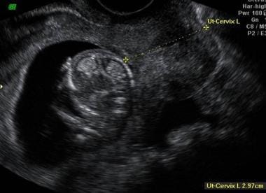
baseline vaginal ultrasound
Sometimes a stubborn corpus luteum cyst sticks around even after your period starts. This procedure is used to ensure your ovaries are not producing eggs at the moment are suppressed.
Sonographic Characterization And Surveillance Of Paravaginal Smooth Muscle Tumor Of Uncertain Malignant Potential Zamora Journal Of Clinical Ultrasound Wiley Online Library
A transvaginal ultrasound can show abnormal structures or growths in your pelvic area that may indicate a condition or disease.

. In addition we provide thyroid abdominal DVT pelvic and renal ultrasound tests to help diagnose and treat other womens health concerns. The first evaluation done on a female is a 3D 4D baseline transvaginal ultrasound USG. Glen Site 1001 Decarie Blvd.
Our Hologic Digital Bone Scanner uses a unique pencil beam targeted X-ray which. Obstetrics and Gynecology Ultrasound 6th floor of Block C Room C 064351. If you are undergoing infertility treatment you will have a baseline ultrasound where we check to confirm that your ovaries have not formed any unexpected cysts that may disrupt your treatment.
The ultrasound is trans-vaginal a wand-like transducer is inserted vaginally to look at your ovaries uterus and endometrial lining. We also look at the endometrial lining thickness to make sure it. The purpose is to check that there are no unusual cysts on the ovaries before starting the fertility drugs.
Ultrasound remains one of the most routinely performed medical investigations and a mainstay of clinical decision-making rests upon the obtained results. This non-invasive test is performed using advanced DEXA equipment to identify potential bone mineral loss or osteoporosis. For more information about transvaginal ultrasound.
These organs include your cervix uterus fallopian tubes and ovaries. A few days after your period starts either following priming or normal start of your period without priming you will go to your fertility clinic for a baseline vaginal ultrasound and blood tests. Iui infertility vaginalultrasoundWatch as I discuss what the baseline ultrasound process was like what the doctor is looking for and what to expect next.
In some cases an endometrial sample or biopsy may be performed. This helps in assessment of the uterus and thickness of endometrium. The ultrasound may take 1530 minutes.
A requisition is needed if you are doing this ultrasound elsewhere. It is also used to diagnose pelvic pain menstrual and gynecological problems abnormal bleeding and certain types of infertility. A baseline ultrasound is a scan done early in your menstrual cycle before starting any fertility medications.
This ultrasound examination also gives us a sonographic baseline for future. Your doctor will insert a speculum just like for a Pap test and cleanse the cervix with a betadyne soap solution. Call 514 843-1650 on day 1 of your cycle to request an appointment.
Baseline ultrasound Around the time of your expected period we will perform a transvaginal ultrasound scan to examine your ovaries. This is known as your baseline ultrasound. A transvaginal ultrasound is an imaging procedure that allows your provider to see your pelvic cavity and the organs inside your pelvis.
Transvaginal meaning across or through the vagina ultrasound uses sound waves to create images of the vagina cervix uterus and ovaries. A thin catheter tube will then be inserted into your cervical canal to enter the uterine cavity. If an appointment is not available wait for your next cycle.
These tests ensure that you dont have any cysts in your ovaries the lining of your uterus is thin and to confirm that. After the ultrasound is complete the vaginal probe will be removed. Ultrasound exams of your ovaries and uterus will be done two to five times during your treatment cycle depending on your response to.
The goal of this scan is to check how many antral follicles you have in each ovary and to ensure there are no abnormalities in your uterine cavity endometrial lining or ovaries. A transvaginal ultrasound is a safe scan with no known risks. Before I started these drugs my first ultrasound 2 years ago showed my uterus to be in perfect condition.
If you are or have been on Tamoxifen Armidex Anastrozole or Femara it is important that you get a baseline ultrasound of your uterus and then follow up with a second ultrasound two years later. People might experience some light discomfort during it but this should go away afterward. The baseline ultrasound should be done on day two or day three of your period.
The baseline vaginal ultrasound is performed at the. Due to the relative proximity of the pelvic organs to the abdominal surface and easy access via the vaginal route gynaecological scanning should be in the armamentarium of every gynaecologist.
Basic Sciences In Gynaecology Section 1 The Ebcog Postgraduate Textbook Of Obstetrics Gynaecology
Cervical Incompetence Imaging Practice Essentials Magnetic Resonance Imaging Ultrasonography
Transvaginal Ultrasound B Mode And Color Doppler Results Download Table
Basic Sciences In Gynaecology Section 1 The Ebcog Postgraduate Textbook Of Obstetrics Gynaecology
Basic Sciences In Gynaecology Section 1 The Ebcog Postgraduate Textbook Of Obstetrics Gynaecology
Cervical Incompetence Imaging Practice Essentials Magnetic Resonance Imaging Ultrasonography
Evaluation Of Uterine Fibroids Using Two Dimensional And Three Dimensional Ultrasonography Chapter 2 Modern Management Of Uterine Fibroids
Basic Sciences In Gynaecology Section 1 The Ebcog Postgraduate Textbook Of Obstetrics Gynaecology
Cervical Incompetence Imaging Practice Essentials Magnetic Resonance Imaging Ultrasonography
Cervical Incompetence Imaging Practice Essentials Magnetic Resonance Imaging Ultrasonography
452 Sonographic Predictors Of Antepartum Bleeding In Placenta Previa American Journal Of Obstetrics Gynecology
Cervical Incompetence Imaging Practice Essentials Magnetic Resonance Imaging Ultrasonography
Basic Sciences In Gynaecology Section 1 The Ebcog Postgraduate Textbook Of Obstetrics Gynaecology
Diagnostics Free Full Text Common And Uncommon Errors In Emergency Ultrasound Html
Evaluation Of Uterine Fibroids Using Two Dimensional And Three Dimensional Ultrasonography Chapter 2 Modern Management Of Uterine Fibroids
2d And 3d Ultrasound A 2d Ultrasound In Midsagittal Plane Left And Download Scientific Diagram
Cervical Incompetence Imaging Practice Essentials Magnetic Resonance Imaging Ultrasonography
Transcervical Fibroid Ablation Tfa With The Sonata System Updated Review Of A New Paradigm For Myoma Treatment Springerlink
Comments
Post a Comment